Download presentation
Presentation is loading. Please wait.

1
The Knee Chapter 18

2
Knee Bony Anatomy Lateral Medial Femur Condyles Tibia Tibial Plateau
Fibula Patella Largest sesamoid in body Shape of condyles allows femur to roll and spin on flattened top portion of tibia, called tibial plateau

3
Knee Bony Anatomy

5
Patellofemoral Joint Point where patella and femur are connected in the trochlear grove

7
Tibiofemoral Joint Tibia meets with femur Weight-bearing joint
Hinge joint Joint capsule 4 ligaments Motions: Flexion Extension Rotation of tibia on femur

8
Patella Malalignment Deviations
Genu Varum Genu Valgum

9
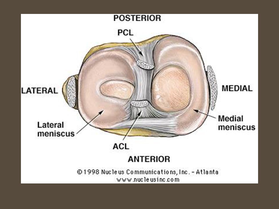
Knee Cartilage & Menisci
Articular cartilage Thin layer of connective tissue over ends of long bones Lateral & Medial meniscus Shock absorption Distribute forces Improve stability of femur as it rides on tibia Synovial membrane Synovial fluid Lubricates articulating surfaces of joints Supplies nutrients to articular cartilage

10
Knee Cartilage & Menisci

11
Menisci Two—medial & lateral Fibrocartilaginous disks
Act as cushions between ends of femur and tibia/fibula Top of tibia flat Condyles of femur rounded Make knee joint more stable

12
Menisci Medial meniscus Lateral meniscus C-shaped
Attached to ligaments on back and medial side of knee Thus does not move freely And torn more often than lateral Lateral meniscus O-shaped Attached only at back of knee Moves more freely as knee flex/extend

15
Ligaments of the Knee Medial Collateral Ligament (MCL)
Lateral Collateral Ligament (LCL) Anterior Cruciate Ligament (ACL) Posterior Cruciate Ligament (PCL)
 Anterior Cruciate Ligament (ACL) Posterior Cruciate Ligament (PCL)")
16
Ligament Attachments Function MCL LCL ACL PCL

21
Muscles of the Knee Quadriceps Hamstrings Vastus medialis
Vastus intermedius Vastus lateralis Rectus femoris Hamstrings Biceps femoris Semitendinosus Semimembranosus

22
Quadriceps Rectus Femoris Vastus Lateralis Vastus Medialis
Extend knee Flex hip Vastus Lateralis Vastus Medialis Vastus Intermedius

23
Hamstrings Biceps Femoris Semitendinosus Semimembranosus Flex knee
Lateral rotate knee Extend hip Semitendinosus Medial rotate knee Semimembranosus

24
Hamstrings

25
Hamstrings

26
Popliteal space

27
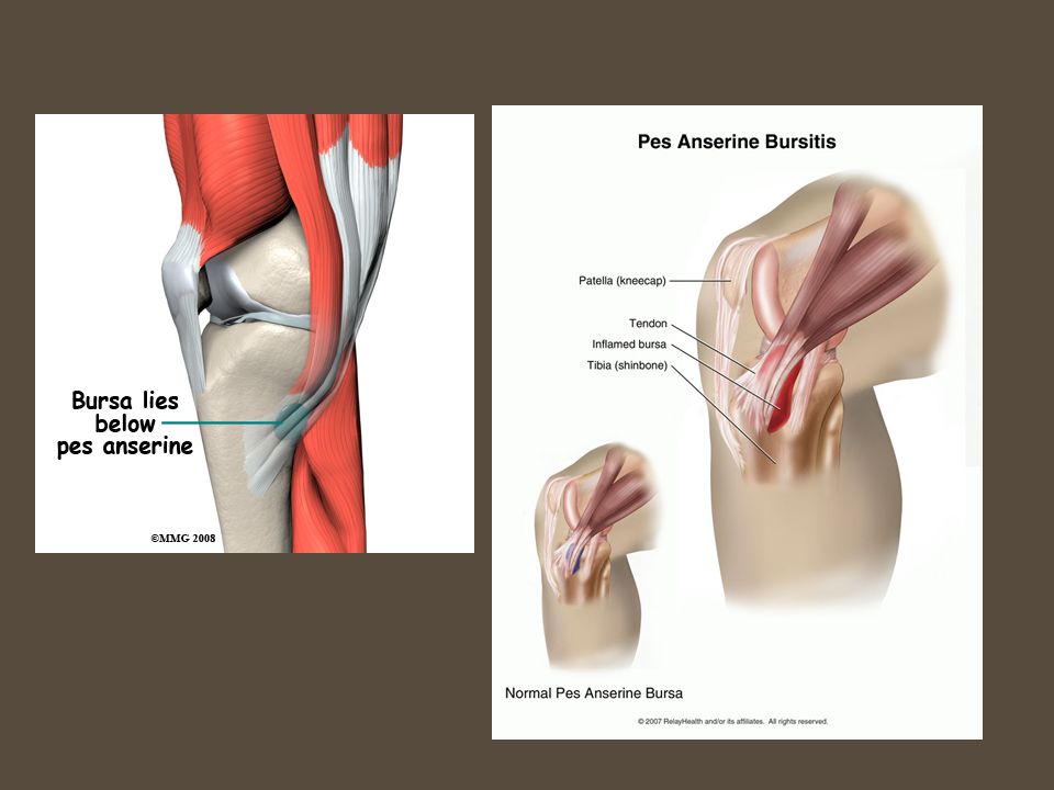
Muscles of the Knee Patellar tendon Sartorius Gracilis Pes Ansurine
Flex hip ER hip Flex knee Gracilis Adduct hip Pes Ansurine

28
Patellar Tendon

29
Gracilis Sartorius

30
Label the Muscles of the Knee

31
Common Knee Injuries

32
Patellofemoral Problems
Symptoms c/o aching pain in front of knee Gradual onset Pain behind kneecap c/o knee giving way Pain going up stairs Crepitus Pain can increase after prolonged knee flexion

33
Patellofemoral Problems
Causes Treatment Femur internally rotated Squinting patella Excessive foot pronation Lowering of the arch Thigh hip internal rotators Weak hip external rotators Orthotics Muscle strengthening Muscle stretching Patellar tracking taping

35
Patellar Tendonitis aka Jumper’s knee
Inflammation of the patellar tendon Signs & Symptoms Anterior knee pain Local tenderness Local swelling Treatment Modify activity Non-impact activities Stretching quads Ice Specialized bracing & taping

36
Fat Pad Syndrome Signs & Symptoms
Inflammation of infrapatellar fat pad Fatty tissue lying deep under patellar tendon Hoffa’s fat pad Often confused with patellar tendonitis Pain just below patella Movement of knee aggravates symptoms Knee tender to palpation Swelling in anterior portion of knee

37
Fat Pad Syndrome—Treatment
Strengthening exercises Avoid full knee extension Leg press Specialized taping Ice NSAIDs

38
Fat Pad Syndrome—Special Test
Pressure applied to proximal patellar tendon with quadriceps contracted Stressing only the tendon and not the fat pad Pressure applied over proximal patellar tendon with relaxed tendon Allow compression of the fat pad

39
MCL Sprain Signs & Symptoms MOI
Pain & tenderness on medial aspect of knee Joint line Bony attachment sites Limited motion in full flexion and extension Swelling Varying degrees of laxity MCL Sprain MOI Valgus force on medial tibiofemoral joint Blow to lateral aspect of knee High-energy twisting maneuver

40
MCL Sprain—Treatment PRICE Rehab Gentle active & passive stretching
P: ace, brace, or crutches Rehab Submax strengthening in subacute stage, but only if painfree Bike once gain flex degrees Gentle active & passive stretching Avoid valgus & twisting forces

41
LCL Sprain Not frequently involved in sports injuries
MOI: varus stress on lateral tibiofemoral joint Signs/symptoms & treatment similar to those of MCL sprain

43
MCL/LCL Sprain—Grade 1 Mild tenderness over ligament
Usually no swelling Pain felt with valgus/varus test but no laxity

44
MCL/LCL Sprain—Grade 2 Significant tenderness over ligament
Some swelling seen over ligament Pain and laxity in joint with stress test, but definite end point

45
MCL/LCL Sprain—Grade 3 Complete tear of ligament Pain can vary
Sometimes not as bad as Grade 2 When knee stressed, definite joint laxity Athlete may c/o knee wobbly or unstable

46
ACL Injuries Females who participate in soccer and basketball 4-6 times more likely than males who play same sport 70% are non-contact injuries Why incidences higher in females?

47
Female Factors & ACL Biomechanical factors Hormonal influences
Use quads more than hamstrings Land on flat foot vs toes Hormonal influences Estrogen levels Environmental factors Anatomic risk factors

48
ACL Tear Contact or non-contact Low to lateral knee
Knee joint in combined position of flexion, valgus, and rotation of tibia on femur Once stretched or ruptured, will not heal Often accompanied by meniscus tears and/or MCL sprains

49
ACL Tear—Signs/Symptoms
Heard or felt “pop” Rapid effusion Knee “buckles” or “gives way” Special testing—Lachman’s or Anterior Drawer Test’s ligaments integrity Within first 5 min to avoid protective muscle guarding Often false-negative testing F/u with orthopedist MRI to confirm

50
Lachman’s Test

51
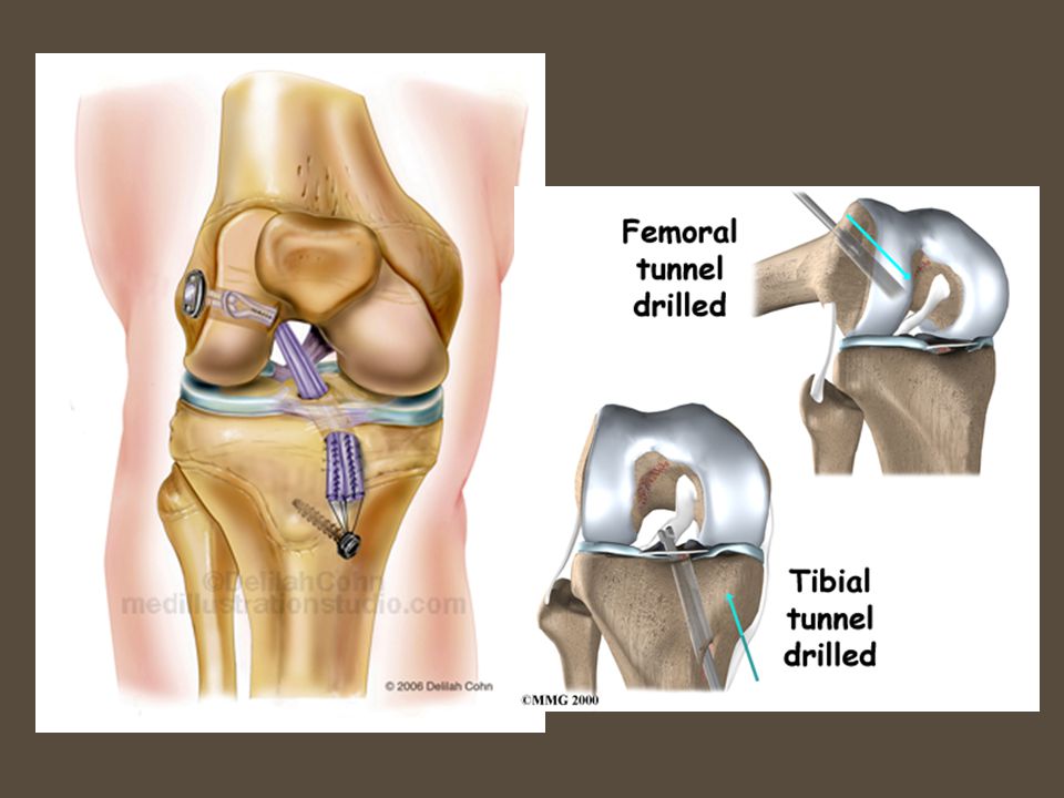
ACL Tear—Treatment Acute: splint, ice, compressive wrap, crutches
Reconstructive surgery necessary to replace ACL Patellar tendon Hamstring tendon (Gold standard) Cadaver Comprehensive rehab (6 months)
 Cadaver. Comprehensive rehab (6 months)")
52
http://physiomed. patientsites
54
PCL Injuries Account for 3-20% of all injuries
Less researched because injured less often (compared to ACL) MOI: tibia strikes ground/object and is pushed backward Motor vehicle accident Industrial accident Fall on flexed knee with foot plantar flexed Hyperflexion of knee
 MOI: tibia strikes ground/object and is pushed backward. Motor vehicle accident. Industrial accident. Fall on flexed knee with foot plantar flexed. Hyperflexion of knee.")
55
PCL Signs & Symptoms Treatment PRICE Rehab Surgery usually avoided
Positive Posterior Drawer Positive Godfrey’s (Sag) Test Athlete in supine position, knee bent at 90⁰ Treatment PRICE Rehab Strength Quadriceps Proprioception Surgery usually avoided
 Test. Athlete in supine position, knee bent at 90⁰. Treatment. PRICE. Rehab. Strength. Quadriceps. Proprioception. Surgery usually avoided.")
56
Meniscus Tears Knee twisted suddenly Ligaments in & around knee torn
One or both menisci become trapped between femur and tibia Ligaments in & around knee torn As ages, menisci lose their rubbery consistencywill soften and fray Weakened structures torn more easily

57
Meniscus Tears

58
Meniscus Tears Treatment Ice Compressive wrap/knee support
Crutches prn Rehab (non-surgical) Streength ROM Activity modification NSAIDS Support sleeve Surgery MRI Signs & Symptoms Mild knee swelling over several hours or more Pain Popping Locking Giving way of knee
 Streength. ROM. Activity modification. NSAIDS. Support sleeve. Surgery. MRI. Signs & Symptoms. Mild knee swelling over several hours or more. Pain. Popping. Locking. Giving way of knee.")
59
Meniscus Tears—Special Tests
McMurray’s Apley’s Compression & Distraction

60
Epiphyseal (Growth Plate) Injuries
 Injuries")
61
Epiphyseal (Growth Plate) Injuries
 Injuries")
62
Osgood-Schlatter Group of symptoms involving the tibial tubercle epiphysis Tibial tubercle: small bump on tibia where patellar tendon attaches Condition result of traction Femur growing faster than quadriceps muscle Result: quad will exert undue pressure on growth center of tibia (at tubercle) Most likely affect males yo and females yo
 Most likely affect males yo and females yo.")
63
Osgood-Schlatter

64
Osgood-Schlatter—Signs & Symptoms
Pain over tibial tubercle Swelling over tibial tubercle Weakness in quad muscles Increased pain & swelling with activity Visible lump Point tenderness over affected area Susceptible to avulsion fx

65
Osgood-Schlatter—Treatment
Address pain, swelling, flexibility During practice/competitionwear protective padding Volleyball knee pad Combine with neoprene sleeve After activityice (even if not painful) & NSAIDs Hamstring tightness cause quads to pull harder during athletic activity Avoid quad stretching (or try gentle stretch) Limit or restrict activity (decrease intensity)
 & NSAIDs. Hamstring tightness cause quads to pull harder during athletic activity. Avoid quad stretching (or try gentle stretch) Limit or restrict activity (decrease intensity)")
66
Osgood-Schlatter—Rehab
Exercises to minimize strength loss rather than increase strength SLR Body weight squats Hamstring curls Calf raises AVOID: Knee extension Heavy squats Power cleans Plyometrics Maintain aerobic fitness Cycling Slide board Swimming

67
Iliotibial Band Syndrome
Irritation usually at femoral lateral epicondyle Bursa facilitate smooth gliding motion of ITB, when inflamed ITB not glide easily Pain worsens with continued movement

68
Iliotibial Band Syndrome
Sudden increase in activity level i.e. runners who increase mileage Mechanical problems: Over-pronate Leg-length discrepancies Bowlegged

69
Iliotibial Band Syndrome
Treatment Gait analysis Review training program Proper footwear Ice Stretch Modify training program Reduce activity level Cross-train Special Test Ober’s

70
Fractures High-energy trauma Rare in athletes
Fx to patella result of direct impact to anterior knee Distal femoral & proximal tibial fxs occur from violent twisting injuries Fall from height (pole vaulter who misses landing pit)
")
71
Patella Dislocation MOI Signs/Symptoms
Plants foot, decelerates, Internally Rotates thigh Signs/Symptoms Obvious deformity Pain Swelling Loss of function

72
Patella Dislocation Management Patella apprehension test Gently extend
Immobolize Rule out: osteochondral fx Patella apprehension test

73
Bursitis

75
Rehab Wall squat Step-up Resisted terminal knee extension

Similar presentations